
Spinal Surgeries
Intervertebral disc disease represents the most common cause of spinal cord injury in dogs and can occasionally occur in cats.
Intervertebral Disc Disease (IVDD) is a broad term, widely used by veterinary clinicians, referring to disc degeneration and clinical neurological disease due to disc herniation.
IVDD can lead to Intervertebral disc herniation which consists of the displacement of the Intervertebral Disc (IVD) from its physiological site leading to spinal cord compression and causing severe inflammation and pain.
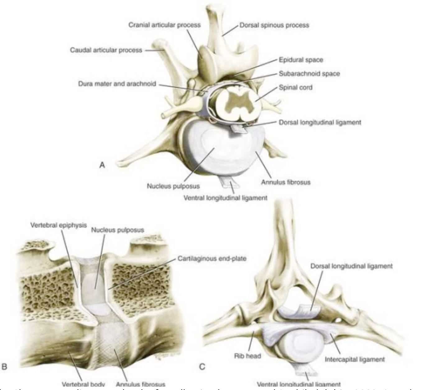
The IVD is a cylinder-shaped structure that lies between adjacent surfaces of the vertebrae. All the IVDs consist of three different anatomical parts: the outer annulus fibrosus, the central nucleus pulposus, and the cartilage end-plate that anchor the disc cranially and caudally to the adjacent vertebral bone. The dynamic interplay of these portions is responsible of the unique functional property of intervertebral discs. They act as viscoelastic hydrodynamic cushions absorbing shocks and providing spinal flexibility.
What causes IVDD?
IVD herniation typically occurs following disc degeneration, although, in rare cases, exogenous trauma can be implicated as well, resulting in the herniation of a non-degenerate IVD.
Disc herniation has been sub-classified as a disc extrusion or Hansen type I, and a disc protrusion or Hansen type II, based on morphology, gross pathology and MRI. Differentiating these subtypes of IVD herniation may lead to different treatment recommendations and outcomes.
HANSEN TYPE I
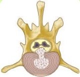
Disc extrusion (Hansen type I) is a type of disc herniation associated with a defect in all layers of the annulus fibrosus which allows displacement of the degenerate nucleus pulposus into the vertebral canal.
It occurs when the disc is no longer able to act as a shock absorber or help with the weight load put on the spine. This happens when the disc undergoes degenerative processes that make it dehydrated. When the IVD degenerates, even normal movements such as jumping or twisting can lead to acute disc herniation. The onset of clinical signs is usually acute.
This process can be age-related but there are certain predisposed breeds that can suffer disc problems from when they are young adult dogs.
 These breeds are referred as ‘chondrodystrophic’ and are characterized by having disproportionably short and curved limbs, such as:
Although the majority of the dogs above are small to medium breeds, larger breeds can also be affected.
 A variant of Hansen type I extrusion is the ‘high-speed low-volume’ (HSLV) or ‘acute non-compressive NP extrusion’ (ANNPE) which occurs when a hydrated and healthy nucleus pulposus is exposed to a sudden increase in intradiscal pressure during vigorous exercise (such as running and jumping) or trauma. The hydrated NP is explosively expelled through a tear in the AF resulting in spinal cord contusion. Then the NP dissipates within the epidural space causing minimal to no spinal cord compression. In both cats and dogs, the incidence of HSLV is low in comparison with the number of patients who have clinical signs referable to IVD degeneration and its consequences.
HANSEN TYPE II
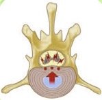
 A Disc Protrusion (Hansen type II) is more similar to human disc disease and consists of a slow protrusion of the disc into the vertebral canal.) It occurs when a rupture affects the inner layers of the annulus fibrosus while the outer layers remain intact and bulging. This event leads to partial displacement of the nucleus pulposus into the disrupted annulus fibrosus.
The onset is usually chronic, with developing clinical signs in weeks to months. This type of IVDD is more common in large non- chondrodystrophic breeds such as German shepherd and Labrador retriever.
Clinical signs associated with IVDD
The severity of clinical signs may vary from pain only to severe neurologic dysfunction, as detailed in the following scale:
Grade I – Spinal hyperesthesia. Ambulatory with no other deficits noted.
Grade II – Tetra or Paraparesis. Ambulatory with loss of proprioceptive function, weakness and ataxia.
Grade III – Tetra or Paraparesis. Non-ambulatory, but evidence of voluntary movement in the limbs.
Grade IV – Tetra or Paraplegia. Non-ambulatory, with no evidence of voluntary movement in the limbs, deep pain sensation present. Urinary incontinence.
Grade V – Paraplegia. As above, with no evidence of deep pain perception.
This grading system is of help in describing the degree of injury; however, as a prognostic indicator, the most significant factor is the presence or absence of deep pain sensation.
Diagnosis
The first step of almost any diagnostic protocol is the signalment, the collection of a thorough history and a complete physical examination. In the investigation of the patient with suspected IVD herniation, a complete neurological examination is carried out with the aims to localise the lesion to specific spinal cord segments and to determinate its severity.
Spinal radiographs may reveal characteristic changes of disc disease such as calcified disc material within the vertebral canal or narrowing of the IVD space or the foramen, however, radiographs rarely provide the accurate conformation and localisation required for surgical management.
For a definitive diagnosis it is necessary to demonstrate spinal cord compression, which can be obtained by CT or MRI. Patients must lie completely still for their scan and this is only possible with the use of sedation (for CT) or general anaesthesia (for MRI).
Prognosis and Treatment of Intervertebral Disc Herniation in dogs
Intervertebral disc herniation, secondary to premature disc degeneration, is a common cause of pain and neurological signs in dogs.
The severity of clinical signs may vary from pain only to severe neurological dysfunction, as detailed in the following scale:
Grade I – No neurological deficits
Grade II – Ataxia/ambulatory paresis
Grade III – Non-ambulatory paresis
Grade IV – Plegia
Grade V – Plegia with no proception
Prognosis and treatment options are grade dependent, as shown on the charts.
Prognosis

Average week of recovery
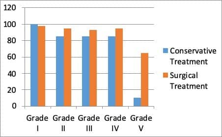
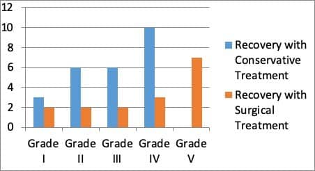
Conservative treatment may be considered in grade I and II cases, although the recovery period is significantly shorter with surgical treatment. In grades II to V the surgical treatment increases the success rate and should be considered. It’s important to note that for Grade V cases surgery should be performed within 48hours of the first clinical signs, after which treatment prognosis decreases to less than 5%.
Contact Northwest Referrals for more information. Tel: 01942 242001 or send us an email at info@northwestreferrals.co.uk.
Hemilaminectomy - (IVDD) - (T1-S1) - £3995
- This includes all consumables, catheterisation, general anaesthetic, hospitalisation for one night (further days are charged at £250/night)
- Plus, a post operative CT scan
Ventral Slot - (IVDD) - (C1-C7) - £3995
- This includes all consumables, catheterisation, general anaesthetic, hospitalisation for one night (further days are charged at £250/night)
- Plus, a post operative CT scan
Please contact us for prices on other neuro surgeries if required.