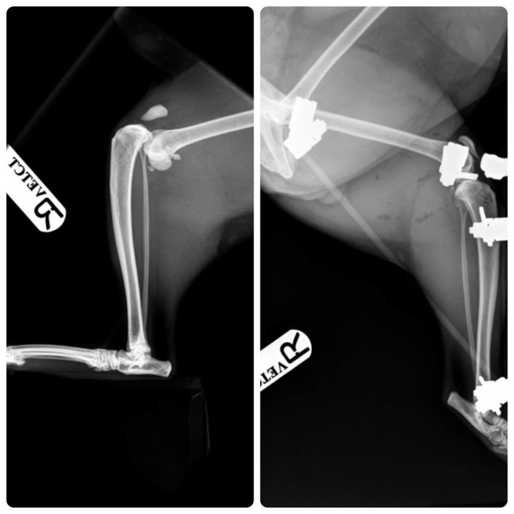
What is External Fixation?
External fixation is a surgical treatment wherein rods are screwed into the bone and exit the body to be attached to a stabilizing structure on the outside of the body. It is an alternative to internal fixation, where the components used to provide stability are positioned entirely within the patient’s body. It is used to stabilize bone and soft tissues at a distance from the operative or injury focus. They provide unobstructed access to the relevant skeletal and soft tissue structures for their initial assessment and also for secondary interventions needed to restore bony continuity and a functional soft tissue cover.
Indications
- Stabilization of severe open fractures
- Stabilization of infected nonunions
- Correction of extremity malalignments and length discrepancies
- Initial stabilization of soft tissue and bony disruption in poly trauma patients (damage control orthopaedics)
- Closed fracture with associated severe soft tissue injuries
- Severely comminuted diaphyseal and periarticular lesions
- Temporary transarticular stabilization of severe soft tissue and ligamentous injuries
- Pelvic ring disruptions
- Certain pediatric fractures
- Osteotomies
- Open fractures that have significant soft tissue disruption (e.g., type II or III open fractures)
- Soft tissue injury (e.g., burns)
- Acetabular and pelvic fractures
- Severely comminuted and unstable fractures
- Fractures that are associated with bony deficits
- Limb-lengthening procedures
- Fractures associated with infection or nonunion
Contraindications
- Patient with a compromised immune system
- Non-compliant owner who would not be able to ensure proper wire and pin care
- Pre-existing internal fixation that prohibits proper wire or pin placement
- Bone pathology precluding pin fixation
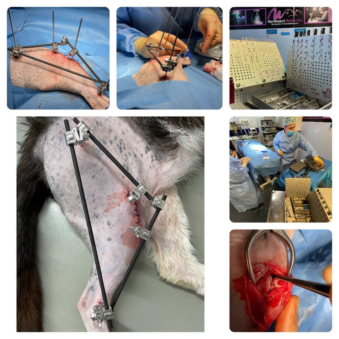
Method
In this kind of reduction, holes are drilled into uninjured areas of bones around the fracture and special bolts or wires are screwed into the holes. Outside the body, a rod or a curved piece of metal with special ball-and-socket joints joins the bolts to make a rigid support. The fracture can be set in the proper anatomical configuration by adjusting the ball-and-socket joints. Since the bolts pierce the skin, proper cleaning to prevent infection at the site of surgery must be performed.
Frame Configuration
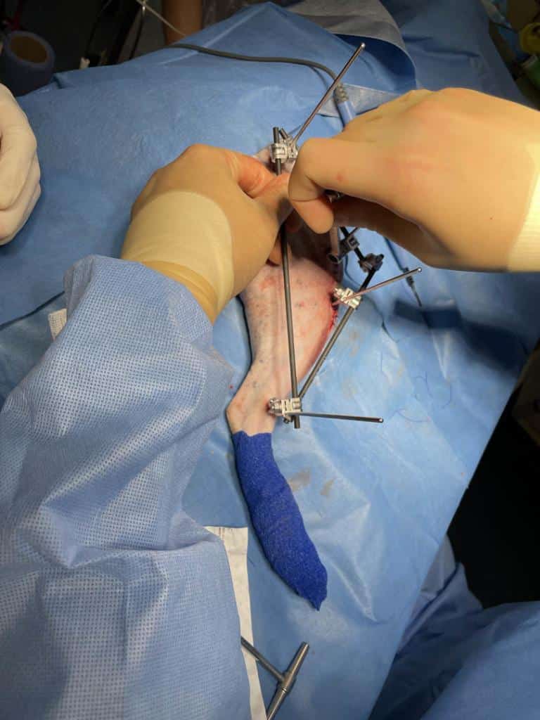
Installation of the external fixator is performed in an operating room, normally under general anesthesia. Removal of the external frame and bolts usually requires special wrenches and can be done with sedation or quick general anaesthetic.
External fixation is usually used when internal fixation is contraindicated- often to treat open fractures, or as a temporary solution.
External fixation is also used in limb lengthening. Typically, the bone is cut diagonally in a surgical procedure. External fixator pins or wires are placed on each side of the split and the external metal apparatus is used to very gradually pull the two sides of the bone apart over a long period of time. Bone will gradually grow into the small gap through distraction osteogenesis.
In most cases, it may be necessary for the external fixator to be in place for many weeks or even months. Most fractures heal in between 6 and 12 weeks. However, in complicated fractures and where there are problems with the healing of the fracture this may take longer. It is known that bearing weight through fracture by walking on it, for example, with the added support of the external fixator frame actually helps fractures to heal.
The parts of an external fixator include:
- Pins ( smooth, negative threaded, positive threaded)
- Connecting rods
- Clamps
Complications
There is a lack of evidence on the best method of pin site dressing to prevent surgical site infections.
History
Almost 2,400 years ago Hippocrates described a form of external fixation to splint a fracture of the tibia. The device consisted of closely fitting proximal and distal Egyptian leather rings connected by four wooden rods from a cornel tree.
In 1840, Jean-Francois Malgaigne described a spike driven into the tibia and held by straps to immobilise a fractured tibia. In 1843 he used a claw-like device to percutaneously hold the fragments of a fractured patella.
Clayton Parkhill of Denver, Colorado and Albine Lambotte of Antwerp, Belgium independently invented the modern concept of unilateral external fixation, in 1894 and 1902, respectively. Lambotte was the first to use threaded pins, however, his device necessitated initial, open fracture reduction and then pin insertion and fixator placement.
In 1938, Raoul Hoffmann of Geneva, Switzerland, building on the work of others, realized that major improvements were essential to making the external fixators more clinically relevant. He developed a technique based on closed reduction with guided percutaneous pin placement. Hoffmann’s technique exemplified the first application of minimally invasive orthopaedic surgery.
In the 1950s, Gavriil Ilizarov of Kurgan, Soviet Union, devised and developed a new method for treating fractures, deformities and other bone defects. A metal frame that encircles the limb is attached to the underlying bone by crossing (X) pins inserted through the bone and limb. The external rings are linked to each other by threaded rods and hinges that allow to move the position of the bone fragments without opening the fracture site, then the fragments can be fixed in a rigid position until complete healing.