Sidney Jenkins, 3y 9m, male Staffordshire Bull Terrier – Lung lobectomy.
Patient X; a 3-year-old male Staffordshire Bull Terrier was referred to NWR with a history of a continuous cough for approximately 8 months following a run out in a corn field.
On clinical examination the patient was otherwise healthy, however, given the history from referring veterinarian our surgeon suspected inhalation of a foreign body.
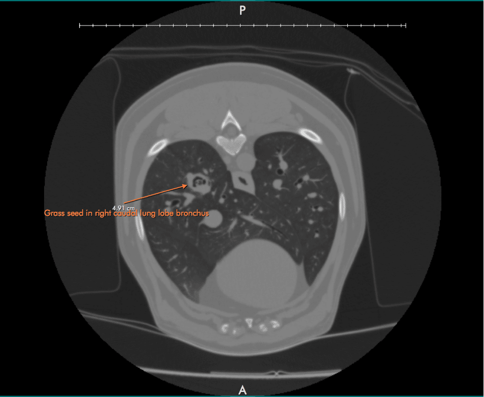
An initial computed tomography was completed in order to evaluate the patient’s thoracic cavity and diagnose the issue. The CT images revealed that a foreign body was present in the caudal principle of the bronchus. This caused oedema, obstruction and bronchesctasia secondary to the initial issue. Despite best efforts the foreign body was unable to be removed endoscopically therefore a surgical approach was discussed.
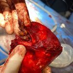
The owners of this patient opted for a lung lobectomy. A lung lobectomy is a surgical procedure that involves partial removal of the lung. Statistics and research show that both dogs and cats can function normally with removal of up to 50% of their lung volume. A right sided intercostal thoracotomy in 6th ICS was performed. A TA stapler was used for lobectomy at the base of the caudal right lung lobe. Once partial removal was complete, evidence of a grass seed was found in the removed lung tissue. The thoracic cavity was flushed with saline and closed using multiple layers of 2 metric vicryl. A Jackson Prat chest drain was placed and in situ for 24 hours post-operative.
The patient is doing great and is expected to make a full recovery.


