 Thomas was an 11yo MN Domestic Shorthair who had been seen several times at the primary practice because he was “snoring, even when he was awake.” He was referred for an MRI to better investigate the increased upper respiratory noise. The referring vet had performed an oral inspection and also a radiographic examination, but no abnormalities had been identified to explain the clinical signs.
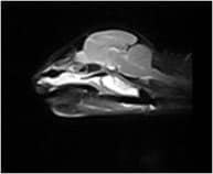
An MRI examination demonstrated a large nasopharyngeal mass resulting in airway occlusion. The lesion is easily visible in the scans shown here but could not be seen on examination. The referring vet was able to incise through the soft palate and remove the lump. Thomas was able to breathe freely for the first time in months! With these unusual cases, early referral for cross-sectional imaging allows you to get to the right diagnosis faster.