
Case 1 – 12yo MN CKCS
 This patient presented to the primary practice with acute neck pain. He could not lift his head above the level of his spine without yelping. He had a previous history of syringomyelia which had been diagnosed on an MRI scan in 2008. Radiographs showed nothing and so he was referred for a repeat scan – the referring vet suspected worsening of the disease condition.
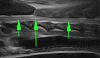
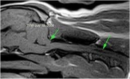
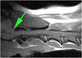 The MR image showed increased T2 hyperintensity and mild widening of the central canal over C2, C3 and C5 (Fig.1, green arrows). There is mild beaking of the cerebellum through the foramen magnum (Fig.2). However, in comparison with the scan from 2008 (Fig.3), the pathology remained largely unchanged.
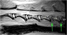
 The scan also revealed that the dog had reduced T2 hyperintensity of the intervertebral disks at C5/C6 and C6/C7 (Fig.4). The disk at C5/C6 is mildly protruding into the vertebral canal and obliterating the ventral epidural fat.
 The owner opted for conservative management of the intervertebral disk disease with non-steroidal anti-inflammatory drugs. The dog significantly improved. This case is a good illustration of two central nervous system pathologies for which MRI scanning gives excellent diagnostic imaging results.