
 This rabbit presented to an exotics specialist following acute paralysis of the hindlimbs. There was no history of trauma and no other obvious clinical signs. The first vet had performed a lateral radiograph but this was non-diagnostic. A spinal lesion was suspected and MRI was the next step.
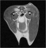 The MRI images showed an abnormally hyperintense signal on the right sublumbar muscles, extensive osteophytosis and osteolysis on the right side of the L3 vertebral body and severe cord compression, as shown on the transverse image on the left.
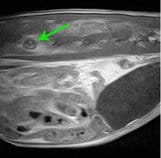
In addition, there was a soft tissue mass dorsal to the caudal bladder, as you can see on the image on the left. Looking at the sequence, this large soft tissue mass bifurcated into two tubular structures.
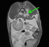
 The sagittal section on the right shows the hyperintense signal under L3 but also shows the soft tissue mass dorsal to the caudal aspect of the bladder. These images are after gadolinium administration.
The report suggested that this was most likely a uterine adenocarcinoma, taking into account the tissue structure and the species. The spinal lesion was likely to be metastatic disease to the soft tissue and bone around L3/L4. Other differential diagnoses were a primary tumour of the sublumbar muscle or extensive myositis and osteomyelitis. The excellent soft tissue detail of MRI was invaluable to the diagnosis of this case.