Phil; a 4 year old, male, cocker spaniel, was referred to Northwest Referrals with a painful neck. On examination his general mobility was within normal limits however this pain resulted in stiffness and reluctance to move the neck.
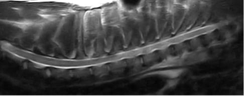
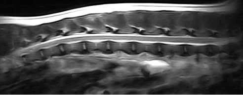
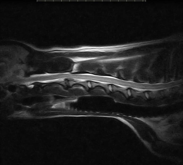
After consultation with surgeon Dr Adam Kluczny MRCVS GP PgC (SAS) a full work up included a neurological examination and MRI scan of the cervical spine to assess the spinal cord in fine detail. The result of this scan would rule out any potential nerve damage, disc disease and tumours, all of which could be the cause of this pain.
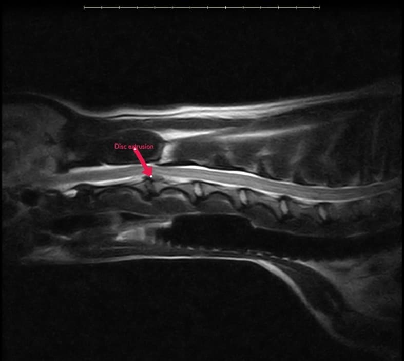
With the MRI scan complete, Dr Kluczny could confirm a diagnosis of a disc extrusion and proposed a plan of surgical decompression of the spinal cord. Phil was then transferred to our surgical team where he was prepared for his surgical decompression.
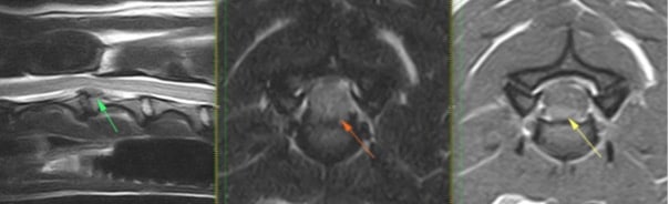
With the assistance of Dr Emily Long BVM BVS BVMedSci MRCVS, a successful surgical decompression of spinal cord in C2-C3 space was complete. This involved removing the extruded disc material that was pressing on the spinal cord. To access this disc space a ventral slot approach was used; skin incision in ventral midline from manubrium sterni to mandible. In addition to this, fenestration of the adjacent discs; C3-C5 was performed. This technique involves removal of the central portion of the intervertebral disc where the nucleus can be found. Recent studies have shown that this technique can reduce the rate of recurrence of disc herniation.
With surgery complete, Phil was then transferred to our inpatient care team for recovery and a post-operative plan was put into place.
Phil stayed with us for one night before heading home and back under the supervision of his primary care vets. His post-operative care plan consisted of pain relief and cage rest for 4 weeks before increasing his exercise weekly. We are absolutely delighted to say that Phil has made a full recovery and is back to his usual, bouncy self. He has since won 1st place in the local dog agility competition that he competes in.