Dexter Johnson. 4y 4m. Neutered French Bulldog
Patient: a 4-year-old, male, neutered French Bulldog, was referred to Northwest Referrals for a spinal tomography as he was unable to walk.
Patient X was non-ambulatory with reflexes absent in both hind limbs, deep pain however was present in this patient. On clinical examination lumbar palpation revealed pain covering the whole lumbar spine region, extending to the last thoracic vertebrae. In this case the patient had ability to control the bladder and anal sphincter.
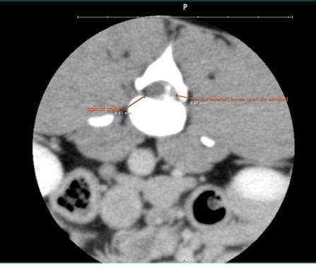
Based upon our clinical examination and referring veterinarian’s assessment, Computed Tomography of the whole spine was done. A Hanson type 1-disc extrusion between L3-L4; affecting the spine up to L1 was discovered. A previous disc extrusion between L5-L6 was also brought to light.
Results from the CT where interpreted and surgery was performed. Patient X underwent a hemilaminectomy extending from L1 to L4 and L3 laminectomies. A hemilaminectomy is a surgical technique where the surgeon partially removes the lamina to decompress the spinal canal. Once removed this allows to spinal canal more room thus relieving pressure off the spinal cord or nerves. Our surgeon used a mid-line, dorsal approach to gain access to the affected areas and in this case extruded disc material. All extruded disc material was removed successfully and defect was closed with 3/0 pds suture. A post-operative Computed Tomography was done to confirm this.
Post-operative the patient cage rest for the next 4-8 weeks with limited exercise to be increased based on patient’s progress. The patient is doing great and is expected to make a full recovery.