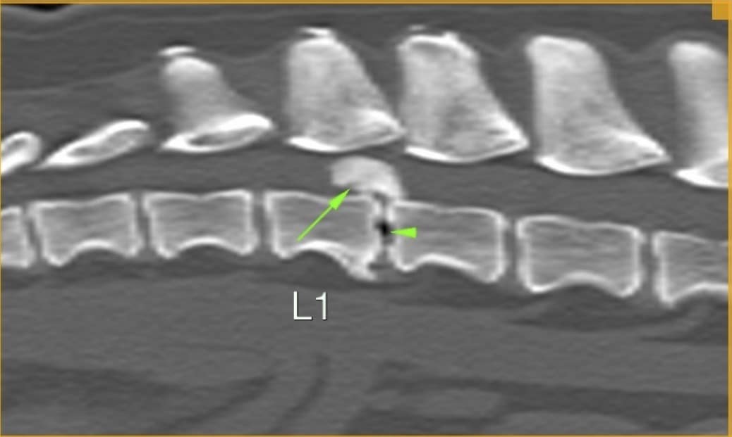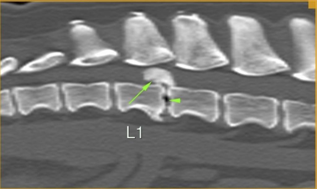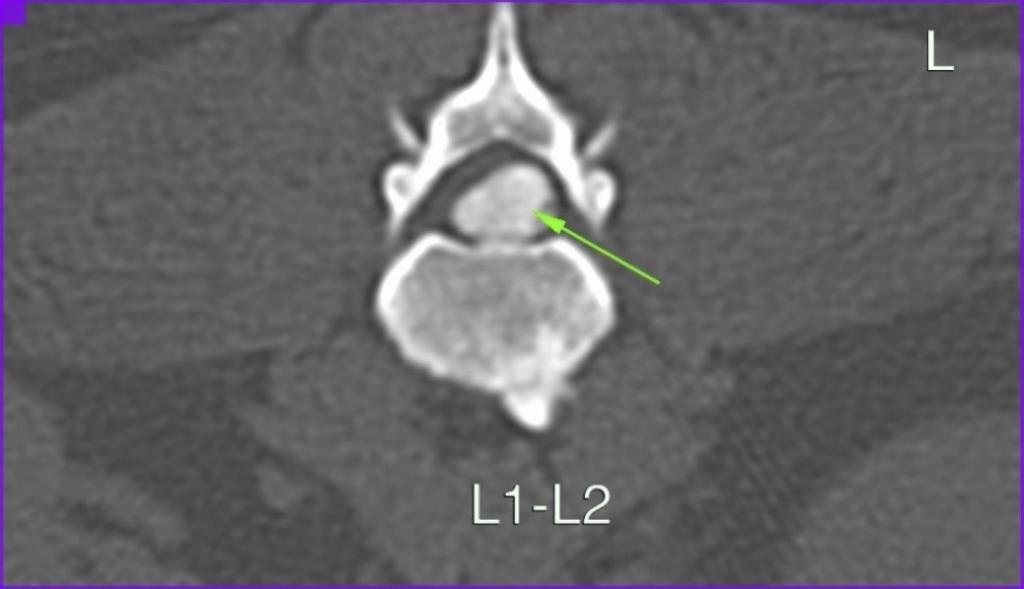
Archie is an 8-year-old, neutered male West Highland Terrier who was referred to the practice for a CT scan. The history from his regular practice disclosed that he started limping on his hind legs two weeks earlier. His symptoms started to get worse despite treatment; Archie began showing acute pain believed to originate from the stomach. He was eventually referred to us when he progressed to hind limb paresis. We performed a spinal CT and found a very large lumbar disc protrusion pressing on Archie’s spinal cord. Even though MRI is a better modality to visualise disc protrusions and their direct and indirect effect on the spinal cord, CT can also be used to successfully diagnose disc protrusions. Surgery by our specialist orthopaedic surgeon was performed to correct this. Archie is track to making a full recovery.