
 Hamish initially presented with a swelling on the side of his chest. The primary practice had performed a fine needle aspirate which revealed pus. The lump was drained and the dog given antibiotics. The lump regressed slightly but then started to grow again.
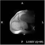
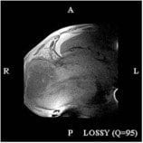 As the dog was so young, the vet opted to refer him for MRI to better evaluate the extent of the disease. Sadly the MRI showed an extremely extensive lesion with invasion into the thoracic cavity and involvement and lysis of six ribs. Often in these types of cases the physical examination only reveals the tip of the iceberg compared to the MRI evaluation.