
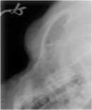
 The first case presented to the referring vet with a small discharging painful lump above the right eye. The dog was clinically normal and the discharge appeared purulent. The dog was initially treated for seven days with clindamycin but the lump remained unchanged. A fine needle aspirate revealed no pus and radiographs showed disruption to the line of the frontal bone. At this point, the dog was sent for an outpatient MRI scan.
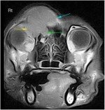 The transverse scan revealed a 4cm mass centred in the right frontal sinus which breached the dorsal portion of the right frontal bone and also crossed over the midline into the left sinus (blue arrow). It also extended into the retrobulbar space (yellow arrow) and into the right nasal cavity (green arrow).
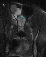 Further caudally, the transverse scan revealed fluid in the right frontal sinus and a breach in the cribriform plate (blue arrow).
Biopsy revealed a squamous cell carcinoma with a very poor prognosis. Surgical removal was considered unfeasible. The owner declined radiotherapy or chemotherapy and the dog was euthanased six weeks after the scan was performed. MRI gives a fantastic resolution of the delicate structures of the head and provides invaluable information for planning surgery and giving prognostic information.